06 Oct, 2025
Feel free to reach out to us.

06 Oct, 2025

This article is medically reviewed by Dr. Sandeep Kumar, Consultant - Surgical Oncology, HCG-Abdur Razzaque Ansari Cancer Hospital, Ranchi.
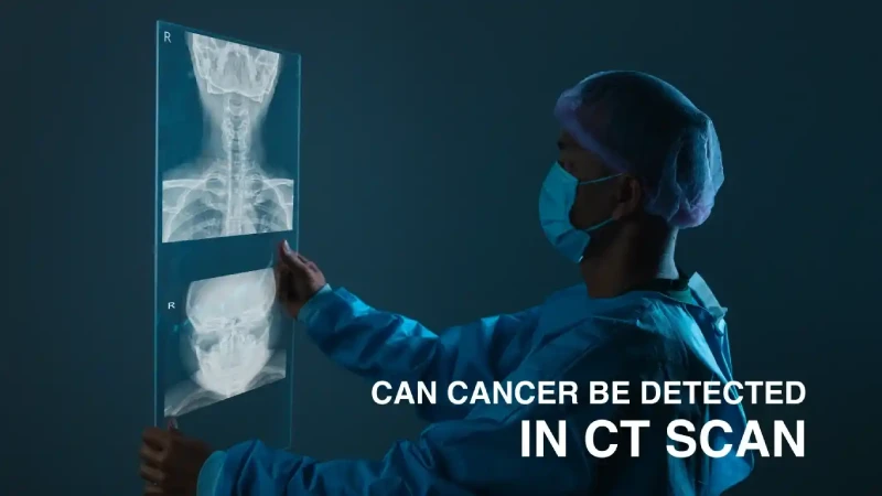
CT imaging has an essential role in cancer care as it helps to detect and stage a tumor and assess how a patient responds to treatment. It is usually performed before surgery to determine the exact location and size of the tumor. CT imaging also plays a role in planning radiotherapy.
A CT scan for cancer provides detailed cross-sectional images of bones, organs, and tissues. Compared to routine X-rays, CT scans provide clearer images and help diagnose cancers.
This imaging method helps determine the site and size of the tumor, along with its relation to adjacent organs and its distance from other organs, which are the key factors considered while planning treatment and determining whether further diagnostic procedures are needed. It may also be done to monitor the effectiveness of treatment during follow-up.
A CT scan for cancer diagnosis is carried out by positioning the patient on a table that moves through a scanner, which captures cross-sectional images of different body parts.
Yes, cancer can be detected in a CT scan. It generates detailed images of the internal organs within the body and helps the physician look for the presence of abnormal growths or tumors. Scans detect cancers even before the symptoms start showing, especially in the lungs, liver, and colons.
CT scan detects the possible spread of cancer to tissues or lymph nodes around the tumor. 3D CT scans can also help assess the size, shape, and location of tumors. Early detection of cancer through CT scans may offer patients several better treatment options and outcomes.
Some of the common types of CT scans are:
A 3D CT scan accurately identifies tumor locations, checks if cancer has spread to other areas, and evaluates treatment effectiveness. It often offers sharper, more detailed images than MRI or ultrasound, with most scans completed in under 10 minutes.
Three-dimensional CT angiography (3D-CTA) is commonly used to diagnose tumors, heart conditions, and other medical issues. This technique provides clear images of small blood vessels and helps in detecting vascular injuries for more accurate diagnoses and improved treatment options.
Results from CT imaging can also help in planning surgeries, especially for treating arteriovenous malformations, as it helps determine the best surgical approach.
Additionally, 3D-CTA is often used as a first-step screening tool instead of digital subtraction angiography to rule out vascular diseases in cases of unexplained spontaneous subarachnoid hemorrhage.
This medical imaging tool creates detailed internal images to assist in planning radiation therapy. It uses simulation, fluoroscopy, and respiratory gating to enable precise radiation targeting.
It locates the abnormalities for guiding radiation therapy, customizes treatment based on the changing breathing patterns of patients, and accommodates different patient sizes and positions. Each session typically takes 15 to 30 minutes, including the time needed for patient positioning and equipment setup.
Multidetector CT (MDCT) is a major advancement in CT scanning technology. Unlike traditional cross-sectional CT, MDCT allows for detailed 3D imaging, making it possible to view any angle and create high-quality 3D visuals.
MDCT scanners perform much faster and capture more extended scan areas or thinner slices as needed. Thanks to its speed and ability to capture fine details, MDCT provides clear, accurate images and significantly improves scan quality compared to older single-detector CT scanners.
PET-CT is not a type of CT scan. However, it is a combination of PET and CT imaging to give detailed information about internal structures and bodily functions. The CT scan produces 3D images of the internal structures, while the PET scan uses a radioactive tracer to highlight highly active cells.
This scan, typically done as an outpatient procedure, takes 30–60 minutes and is more accurate for diagnosing cancer than either scan alone. PET-CT scans help diagnose, stage, plan, and assess treatment effectiveness. They also reveal if tissue that appears abnormal post-treatment is active cancer or just scar tissue.
CT scans are commonly used to diagnose different types of cancer. In rare cases, it may also be used to screen high-risk individuals for cancer. For instance, a CT scan for lung cancer is recommended for people who have been smoking for more than 20 years or have a family history of lung cancer and fall under the high-risk category.
Since a CT scan for cancer provides a clearer view than an X-ray, it is easy to distinguish tumors from other lung tissues. This is critical for accurate and timely diagnosis of lung cancer.
Another instance would be that of breast cancer. A CT scan for breast cancer can help determine the extent of the disease’s spread to different parts of the body.
Other cancer types that can be detected or diagnosed with CT imaging include kidney cancer, bladder cancer, ovarian cancer, colon cancer, stomach cancer, and more.
Few cancers are difficult to detect through CT imaging, and in such cases, other tests, such as MRI, PET scan, or biopsy, will be recommended. Cancers that cannot be diagnosed through CT scan include prostate cancer, uterine cancer, certain liver cancers, certain brain cancers, blood cancer, and bone cancers. For these cancers, CT imaging may be recommended to determine the extent of the disease’s spread (metastasis).
Cancers that are in very early stages cannot be detected by CT scans. Especially in cancers like ovarian, liver, or prostate, small lesions are not easy to identify.
As imaging depends on contrast in tissues, cancers occurring in soft tissues with low contrast appear to be less clear and can lead to a missed diagnosis.
For cancers like multiple myeloma, slight changes in the bone marrow often don't appear on a CT scan. In such cases, other tests are recommended to confirm the diagnosis.
A CT scan for cancer can show the presence of a mass but cannot differentiate between a benign or malignant mass. This often leads to unnecessary tests before arriving at a conclusive diagnosis.
CT scans increase radiation exposure; hence, they can only be used occasionally to check patients, especially young patients and those with cancers that need constant monitoring.
Because of the overlapping structures in tissues, false positives or negative results may sometimes occur in CT for cancer.
There is a difference between MRI and CT imaging for cancer.
Imaging technology has significantly advanced in recent years. Tools like CT imaging and MRI evaluate various tumor parameters before surgery. CT scans are quicker and have a high spatial resolution, making them ideal for detecting abdominal and lung cancers, but they lack soft tissue contrast, limiting sensitivity to small tumors.
MRI, on the other hand, provides superior soft tissue contrast, making it ideal for the detection of small lesions and evaluation of soft tissues, especially in the liver and head/neck.
However, MRI is slower and more expensive than CT. In some cases, both scans have similar performances. CT is often the primary choice due to its speed and cost, while MRI is used for detailed tissue evaluation. These imaging tools help improve diagnosis and treatment planning, leading to better patient outcomes.
Positron emission tomography (PET) and computed tomography (CT) scans have an important role in the diagnosis and staging of cancer. A CT scan for cancer provides detailed anatomical images, helping map tumor size and location, while PET scans focus on metabolic activity, distinguishing between benign and malignant cells.
A PET scan for cancer is effective in assessing isolated pulmonary nodules. It detects the cancer that has spread to lymph nodes. Thus, it reduces the need for invasive staging.
Undergoing PET-CT for cancer diagnosis helps doctors obtain both anatomical and metabolic information, improves diagnostic accuracy, and aids in personalized treatment plans. Together, these scans provide a detailed view of cancer. It results in better treatment decisions and outcomes.
Before a CT scan for cancer, you would be asked to remove metal items like jewelry, piercings, underwire bras, dentures, and hair clips that could interfere with imaging. You’ll wear a gown and inform the technologist about any medical devices like pacemakers.
The CT scanner is a doughnut-shaped machine. You'll lie on a flat table that moves through its center while the X-ray tube rotates to create images. The scan is painless, but holding still may be uncomfortable, and you may need to hold your breath briefly. Some scans require a contrast dye, which may cause a warm sensation or metallic taste in the mouth.
Although CT scans are generally safe, there are some risks, especially when contrast dye is used. Some people may experience mild reactions like rash, nausea, itching, or shortness of breath, which typically go away on their own. However, more serious reactions, including difficulty breathing or low blood pressure, are rare and need immediate attention.
The contrast dye can also affect kidney function, particularly in those with existing kidney issues, so doctors may check kidney function before administering it. There is also a small risk of radiation exposure, which slightly increases the long-term cancer risk. Pregnant women should only undergo CT scans in emergency cases.
Early detection of cancer is important in successful treatment, and CT scans have a key role in this. CT scans can spot tumors that might be too small for physical exams or other imaging methods to detect and help in detecting various types of cancer even before they start showing symptoms.
The technique is used to diagnose various serious conditions, such as pulmonary embolism, heart attack, and stroke. Intravascular ultrasound also assists doctors in determining the need for stent placement in a particular patient.For example, colon cancers often begin as tiny polyps that grow over time, making early detection through CT colonography essential. Early detection of cancer, for example, in lung cancer, leads to higher rates of survival and improved outcomes. CT scans help doctors detect abnormalities in organ size, tumors, and other potential signs of cancer. It helps in early intervention.
Advanced imaging technologies like MRI, ultrasound, and blood tests offer better prevention and treatment options. Early cancer diagnosis through CT scans and appropriate interventions has better outcomes and supports a better quality of life.
CT scans aid in cancer diagnosis, staging, treatment planning, and treatment monitoring. It assists in diagnosing various cancers, such as certain abdominal cancers, lung cancer, kidney cancer, ovarian cancer, and pancreatic cancer. However, CT scans have certain disadvantages, such as radiation exposure and the inability to detect cancer at early stages.
At HCG Oncology, which is a leading cancer hospital in India, we have a dedicated radiology and imaging department, which facilitates different types of CT scans for the diagnosis of various cancerous and non-cancerous conditions.

Dr. Sandeep Kumar
Consultant - Surgical Oncology
MBBS, MS (General Surgery), MCh Surgical Oncology (IMS, BHU, Varanasi)
Dr. Sandeep Kumar is a highly experienced and accomplished surgical oncologist, and he can be consulted at HCG - Abdur Razzaque Ansari Cancer Hospital, a leading cancer hospital in Ranchi. He specializes in the management of a wide range of cancers, including hepatopancreaticobiliary, gallbladder, breast and gynecological, thoracic, stomach, colorectal, soft tissue, and head and neck cancers. He is recognized for his technical excellence and compassionate patient care. His treatment approach is to provide a comprehensive, multidisciplinary approach, with an emphasis on the best possible outcome for each patient.
To book an appointment with Dr. Sandeep Kumar, click here.
