14 Apr, 2025
14 Apr, 2025
This article is medically reviewed by Dr. Yogendra Yadav, Consultant - Nuclear Medicine, HCG-Abdur Razzaque Ansari Cancer Hospital, Ranchi.
A Positron Emission Tomography (PET) - Computed Tomography (CT) scan is a medical imaging technique. It combines the advantages of PET and CT scans that complement each other to provide highly precise and superior-quality diagnostic information.
It plays an important role in diagnosing cancer cells and assists the oncologists in monitoring the treatment response. PET-CT scans also help detect several diseases, such as neurological and cardiovascular diseases.
During the PET-CT scan, the patient undergoes PET and CT scans simultaneously. During the scan, the nuclear medicine physician injects a small amount of radioactive glucose analog into the patient's vein and is made to remain still for some time so that the injected glucose is absorbed by tissues in the body. The PET scanner rotates around the patient's body and detects the tissues that have increased glucose uptake.
At the same time, the CT scanner captures 3D images that will carry the anatomical information of the tumor, such as its exact location, shape, size, and extent of the disease’s spread.
The technologists precisely combine the metabolic information obtained from the PET scan and the anatomical information from the CT scan to deliver a comprehensive imaging report.
Additional Reading: Here is an article by one of HCG’s nuclear medicine specialists on how PET-CT imaging plays a significant role in cancer management: Role of PET-CT Scan in Managing Cancers | HCG Oncology
Cancer cells are the cells that divide and spread uncontrollably. For division and proliferation, they require more energy compared to healthy cells. Thus, to meet their energy demand, the malignant cells take up a relatively higher amount of glucose.
The PET scan uses the radioactive sugar molecule that is taken up in large amounts by the cancer cells. When the patient undergoes a PET scan after having an injection of the radioactive sugar, the images show brightened sites that may suggest the presence of cancer cells. The PET scan may detect cancers that are difficult to detect with X-ray, CT, or MRI.
The whole-body PET scan for cancer plays an important role in detecting the presence of cancer. Further, it also has a significant role in monitoring the response to cancer treatment.
One of the clinical applications of PET scans is to differentiate between benign and malignant lesions. It helps the oncologists in determining their course of treatment. Further, the PET scan for cancer also assists the oncologists in detecting the stage of the cancer. Information about the stage of cancer helps the oncologists to plan appropriate treatment.
The PET scan also helps in monitoring the response to cancer treatment. Throughout the treatment, the doctor may recommend PET scans to determine the efficacy of treatment and check if there are any changes needed in the treatment plan.
Additional Reading: Interested to know more about how PET-CT imaging plays pivotal role in cancer diagnosis? Read this article by one of HCG’s specialists: PET - CT Scan for Cancer Diagnosis: Procedure and Benefits
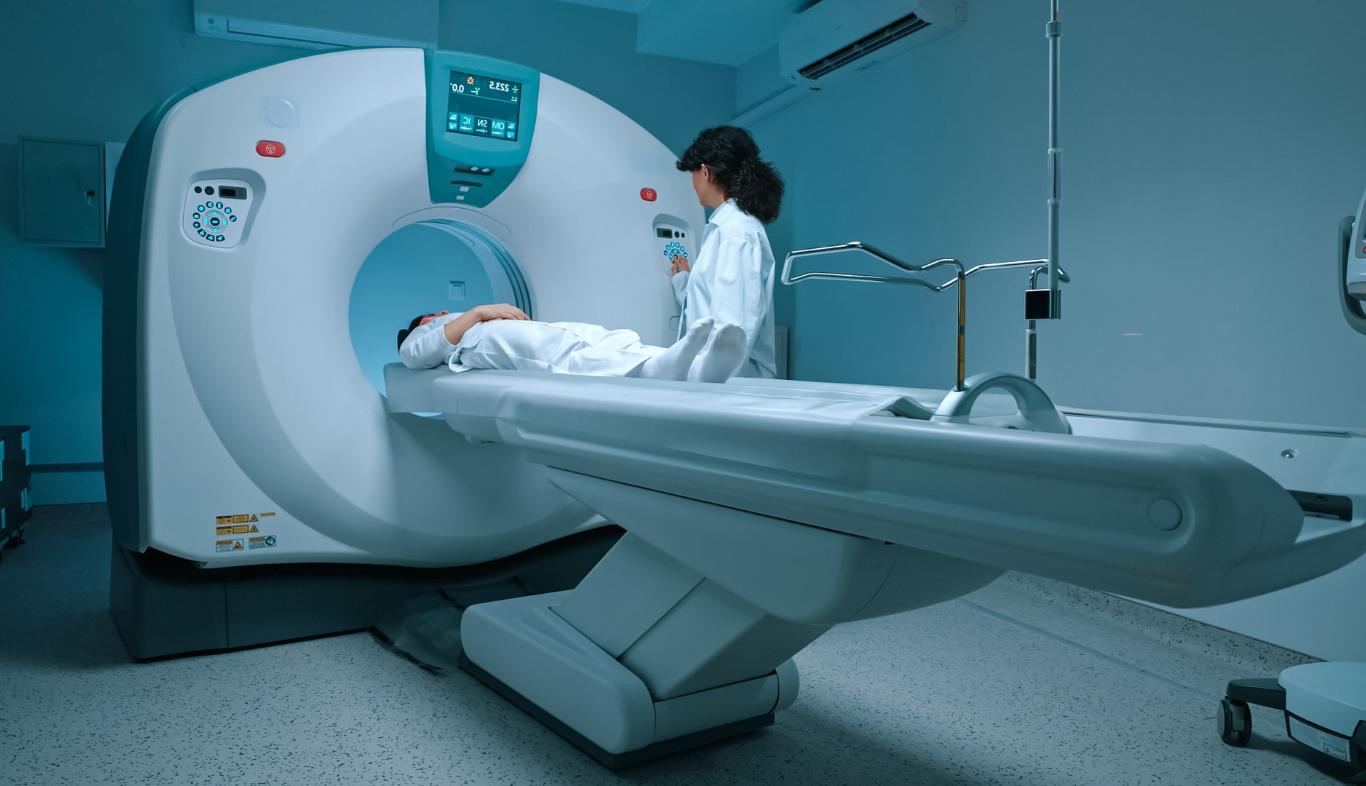
You are taken to the scanning room for a PET scan. The nuclear medicine physician instructs you to lie on the examination table on your back. You may be asked to hold your breath and/or to remain still during the PET scan. Throughout the scan, the NM technologist/physician will be in touch with you through the intercom system.
When the PET scan machine starts, the exam table with you on it slowly moves into the scanner machine. While you move into the machine, the CT scanner captures the images. The nuclear medicine physician reviews the images and shifts you to the waiting room if the images are fine.
The PET test for cancer takes around 1.5 to 2 hours. The maximum time is taken up by waiting after the radiotracer injection to allow the body tissues to absorb the radioactive glucose.
The preparation for a PET scan takes around 15 to 30 minutes; radiotracer uptake takes about 45 to 60 minutes; the actual scanning takes around 20 to 30 minutes; and the post-scan duration is about 5 to 10 minutes. The time also depends on whether it is a whole-body PET-CT scan.
You should follow all the instructions of the nuclear medicine physicians.
Avoid drinking or eating anything at least 6 hours before the examination. You may be allowed to take your medications unless otherwise contraindicated for the scan. The doctor may ask you to arrive 15 to 30 minutes before the scheduled scanning time.
After the scan, the patients are advised to drink an adequate amount of water to flush out the radioactive tracer from the body. Breastfeeding mothers are advised to resume breastfeeding at least 24 hours after the examination.
Inform the doctor immediately if you experience symptoms, such as itchy eyes, nasal congestion, dizziness, vomiting, and restlessness. You may drink or eat normally and perform routine activities.
Arrange for someone to drive you home. Avoid close contact with the babies, pregnant women, or young children for at least 10-12 hours after the examination.
The nuclear medicine physicians combine the results of both the PET and CT scans for interpretation. The images of PET-CT highlight the areas that have high metabolic activity suggestive of inflammation or cancer.
The images also provide anatomical details of the abnormal tissues. Nuclear medicine physicians assess these images to detect abnormalities and to differentiate between benign and cancerous cells.
If the PET scan results suggest the presence of cancer cells, the next step is to confirm the diagnosis of cancer. It is usually done through biopsy. However, oncologists may use PET-CT imaging to determine the disease stage and plan the treatment according to the stage of the disease. The doctors may also monitor the treatment response through PET-CT imaging.
PET-CT scan includes the benefits of both the PET and CT scans. While a PET scan detects the tissues of high metabolic activity, a CT scan detects the exact location of tumors or any abnormal mass. PET-CT scans help in diagnosing various diseases, such as cancer, cardiovascular diseases, and neurological conditions. PET-CT scan is a non-invasive technique with no pain. Like any other imaging method, PET-CT scans also carry a small degree of risk, and therefore, they are only recommended when specialists are sure that their benefits outweigh their risks.
Dr. Yogendra Yadav
Consultant - Nuclear Medicine
Dr. Yogendra Yadav is a highly skilled nuclear medicine specialist who is available for consultations at HCG-Abdur Razzaque Ansari Cancer Hospital, a leading oncology hospital in Ranchi. He has specialized expertise in different nuclear medicine procedures that help in diagnosing and staging different types of cancer. He is known for his patient-focused approach while catering to the needs of his patients.